Gonioscopy, Electroretinography, Ultrasonography
To provide better information on your pet’s condition there are several specialized tools to help aid in diagnosis.
Many pet’s are predisposed to glaucoma due to goniodysgensis. This is a condition in which the drainage angle of the eye was not formed correctly. This angle cannot be seen without the use of a goniolens. Placement of this lens on the surface of the eye, or cornea, allows Dr. Bliss to see any abnormalities. With the information provided from this gonioscopy exam, Dr. Bliss can appropriately direct treatment.
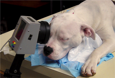
An Electroretinogram (ERG) provides an electrical reading of the fundus, or retina. This test does not measure vision, but measures how well the retina is responding to light. It is usually recommended when a full picture of the fundus cannot be visualized. An ERG aids in the diagnosis of inherited retinal changes, SARDS, as well as consideration for cataract surgery. Pet’s usually need to spend about 20 minutes in the dark before performing this test, and results are available immediately.
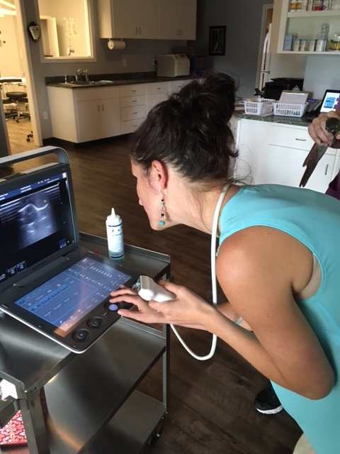
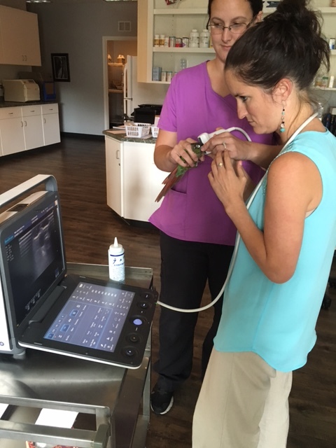
Ultrasonography is a noninvasive way for Dr. Bliss to image and measure the eye. This is usually recommended when certain structures of the eye are difficult to evaluate, have changed in position, or are abnormal. Ultrasounds can aid in the location of intraocular or retrobulbar masses, measuring of the lens for cataract surgery, measuring corneal thickness, and location of the fundus. Most pets accept this procedure with just a topical anesthetic.