“PHOENIX” THE GOLDEN EAGLE TO RECEIVE RARE ARTIFICIAL LENS TRANSPLANT

“Phoenix” the Golden Eagle Eye Cataract and Lens Transplant Surgery

Wednesday, February 3, 2016

Southern Oregon Veterinary Specialty Center, 3265 Biddle Road, Medford


10:00 a.m. Pre-Surgery Media Interviews, Photos, Video Footage with Dr. Bliss and Executive Director, David Siddon

11:00 a.m. Surgery* (Surgery may take up to one hour)

11:30-Noon Post-operative interviews with Dr. Bliss


*Media may film and take photos from window outside of surgery suite


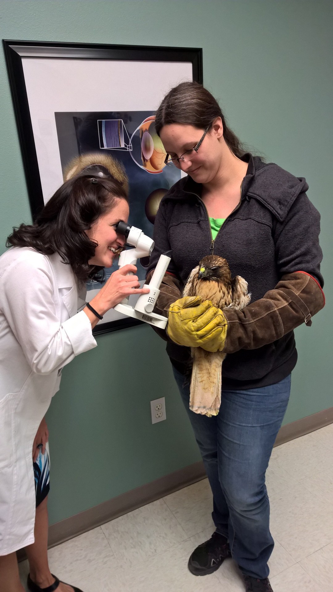
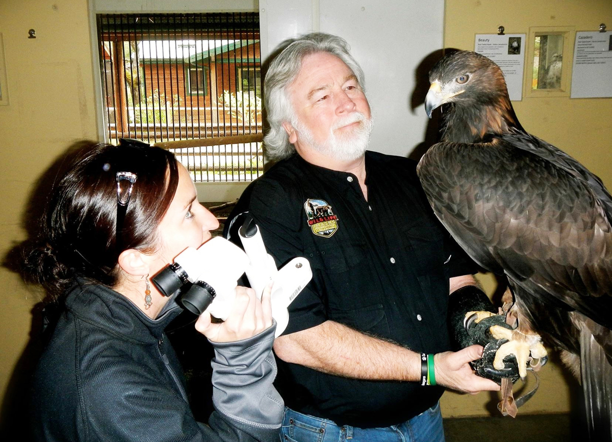
Medford, OR— An appearance in a Disney movie and filming with John Denver won’t be the only reasons “Phoenix” the Golden Eagle has earned some fame. This notable golden eagle that calls Wildlife Images home will be receiving a rare cataract surgery and artificial lens transplant on Wednesday, February 3, 11 a.m. at Southern Oregon Veterinary Specialty Center in Medford. The surgery will be performed by animal specialty ophthalmologist, Dr. Cassandra Bliss.

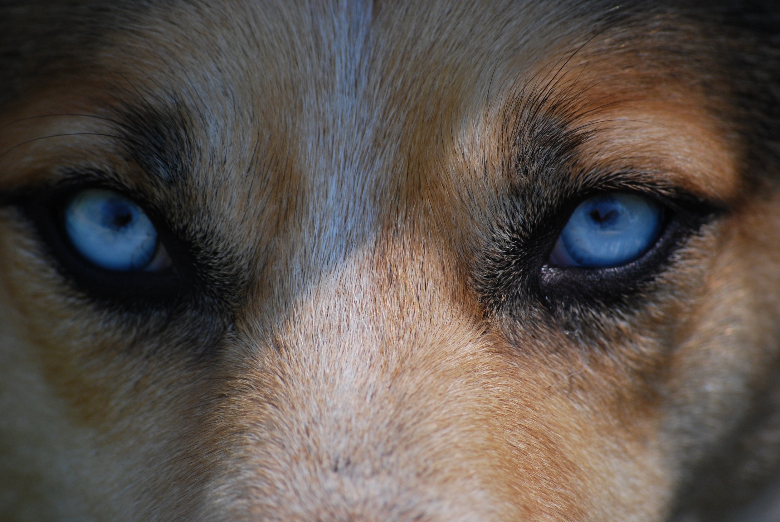
“Thanks to the generous donation of the artificial lens by I-MED Animal Health, we will be able to increase Phoenix’s visibility dramatically,” says Dr. Bliss. “His cataract has progressed to the point where he has nearly no sight in his left eye.” Although this won’t be Dr. Bliss’ first specialized cataract surgery and artificial lens transplant, it will be her first on an eagle. Dr. Bliss has performed this unique and highly specialized surgery on multiple dogs, penguins and other exotic species. “I look forward to helping Phoenix get his vision back,” comments Dr. Bliss.

“Phoenix” the Golden Eagle has a special legacy at Wildlife Images. In 1980, Phoenix arrived at the center as a fuzzy baby eaglet. He had fallen from his nest onto a remote logging road out of Brookings, Oregon. Found by a caring truck driver who tucked him into his hardhat and contacted Wildlife Images, Phoenix began his rescue journey.  As he grew into adulthood, Phoenix was deemed “non-releasable” because of his dependence on humans and in particular Dave Siddon, Executive Director of Wildlife Images. For nearly 35 years Phoenix has resided at Wildlife Images and contributed significantly to the centers educational and outreach programs by providing a deeper appreciation of his species and the importance of conserving and protecting our natural resources. “I’m very thankful to Dr. Bliss for the opportunity to help Phoenix,” said Dave, “what’s important to understand is when an eagle like Phoenix can’t see, his comfort and quality of life can deteriorate rapidly – we’ve noticed this happening in the last few months. The surgery, will give Phoenix many more years of ambassadorship as his life expectancy in captivity can be well into his 40’s.”

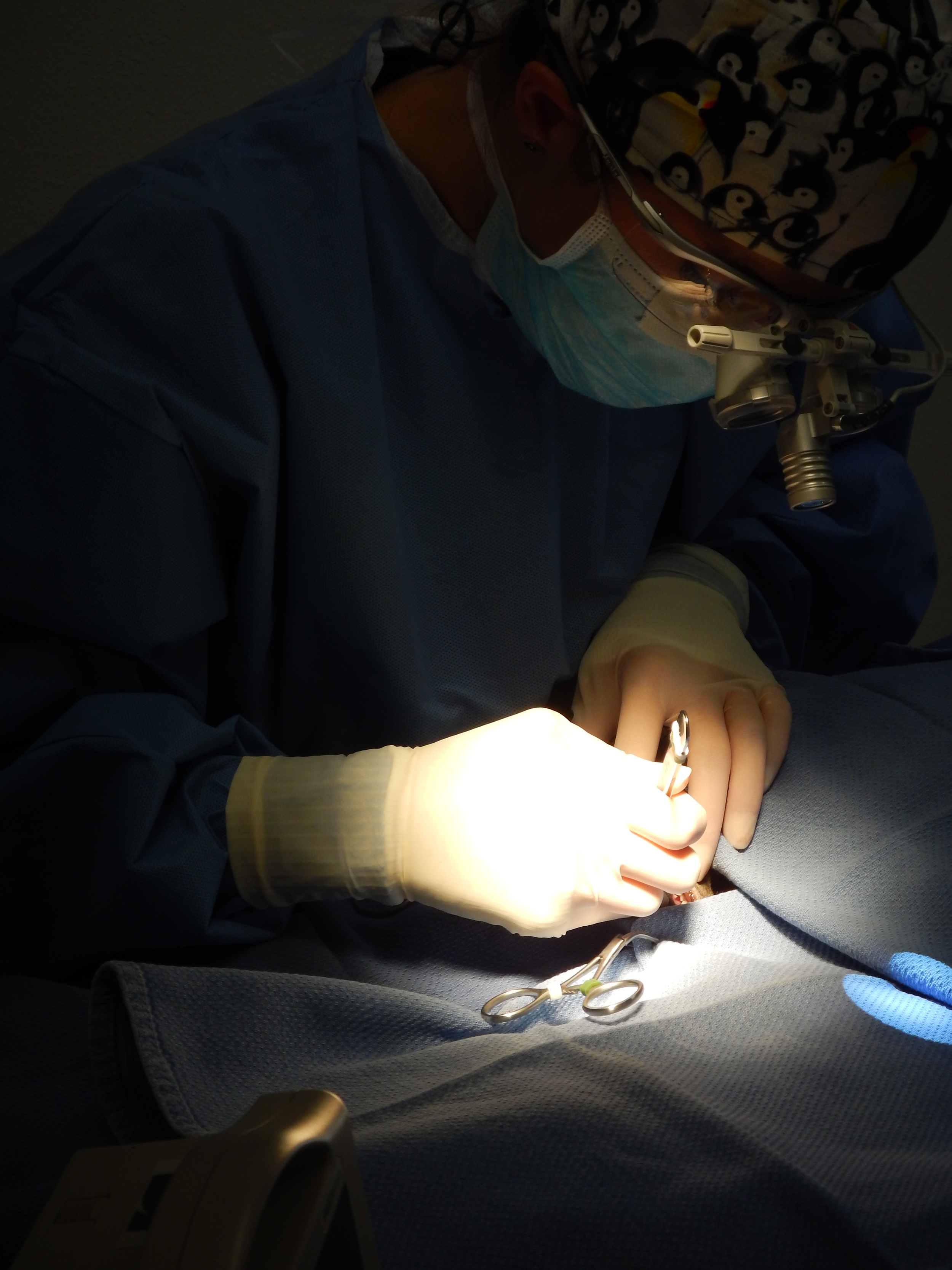
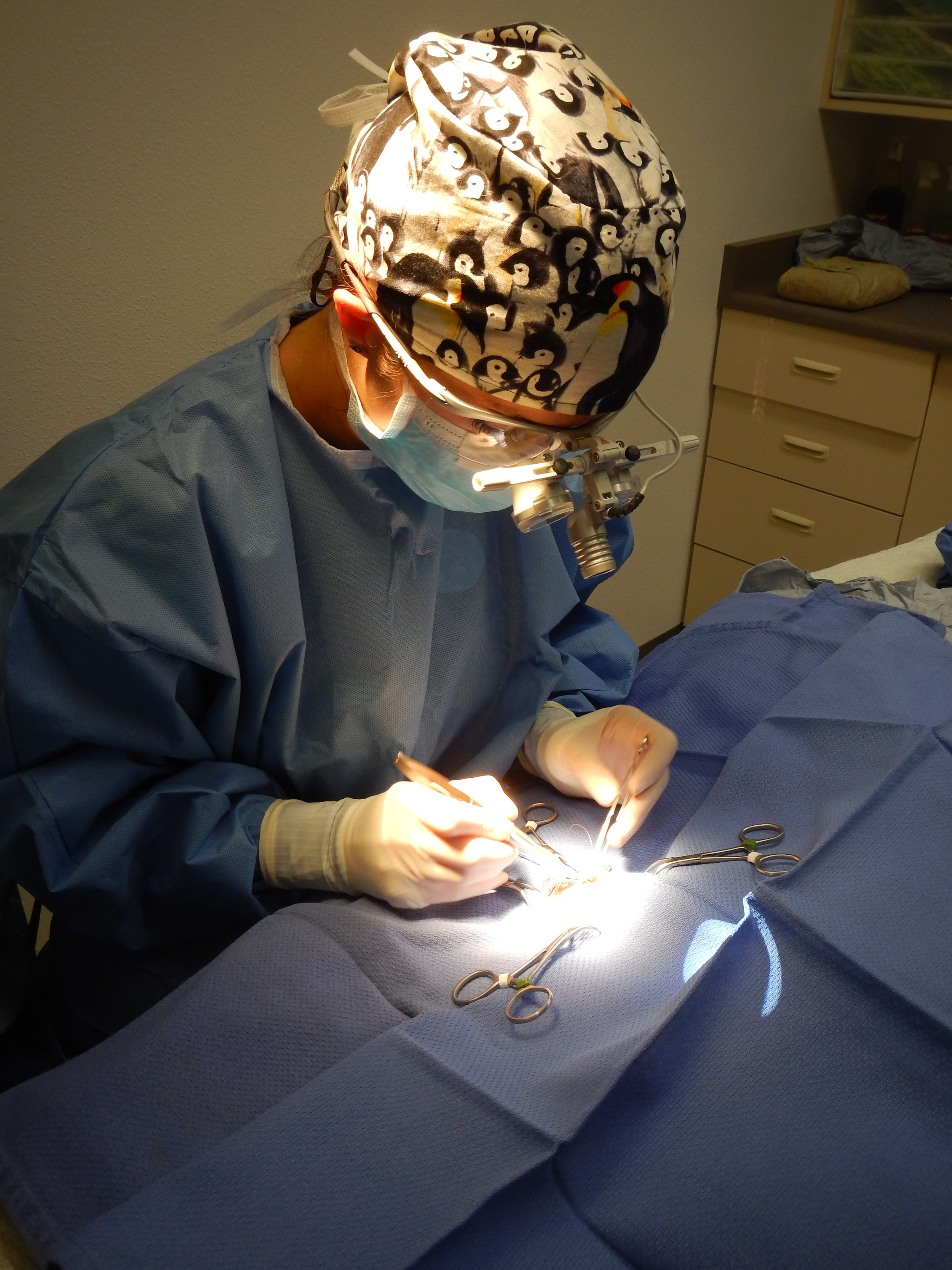
Dr. Bliss is a Diplomate of the American College of Veterinary Ophthalmologists, specializing in cataract and microsurgery, eyelid reconstruction and blepharoplasty, corneal repair and grafting, keratoconjunctivitis sicca and tear film management, glaucoma, equine corneal disease and recurrent uveitis. She has been practicing with Southern Oregon Veterinary Specialty Center since November 2014 and operates her own practice, Bliss Animal Eye Care.

Wildlife Images Rehabilitation and Education Center is a 501(c)(3) non-profit organization established in 1981. They provide care and treatment for sick, injured, and orphaned wildlife indigenous to the Pacific Northwest, while giving the public an opportunity to experience wildlife first-hand. Wildlife Images receives and cares for over 1,000 animals every year at no charge to individuals or organizations. Wildlife Images is a sanctuary for over 80 permanent resident animal ambassadors such as grizzly bears, black bears, cougars, bobcats, badgers, and variety of birds of prey. The center offers extensive outreach and educational programs supporting their core purpose to involve, educate, and inspire children and adults about the importance of preserving and protecting their natural resources while fulfilling their mission of SAVING WILDLIFE. With over 80 resident animal ambassadors at the center, Wildlife Images does not receive any state or federal funding and their survival is based solely on donations from sponsors and donors.

I-MED Animal Health, a division of I-MED Pharma, Inc., is dedicated to improving eye health for all animals.  I-MED Animal Health specializes in surgical and non-surgical products that alleviate Cataracts, Dry eye (KCS), tear stains and other eye conditions.

In collaboration with ophthalmologists and engineers, I-MED was the first company to design replacement lenses for birds of prey. For more information about I-MED Animal Health, visit www.imedanimalhealth.com.

For more information about Southern Oregon Veterinary Specialty Center, please contact Dr. Diana Schropp at 541-282-7711 or visit www.sovsc.com. For information about Bliss Animal Eye Care, visit www.blissanimaleyecare.com.

For more information about Wildlife Images, visit www.wildlifeimages.org. 541.476.0222

Red Tail Hawk Release Back Into the Wild Today at Wildlife Images

Wildlife Images rehabilitation and education center received a Red-Tailed Hawk with a severe eye injury and new exactly who to call!

Kendra Romero, Animal Care team leader from Wildlife Images contacted Cassandra Bliss, veterinary ophthalmologist, of Bliss Animal Eye Care in Southern Oregon to exam the bird. After the examination of the Red-tailed Hawk Dr. Bliss concluded the hawk needed emergency eye surgery. The surgery included the stitching of the laceration in the eye to prevent further damage.

After a successful surgery and recovery, the patient was moved to an outdoor enclosure where it could live, and hunt therefore proving it had the strength to be released back into the wild.

May 7th, 2015 the hawk was given a clean bill of health and was released Friday May 8th near Wildlife Images in Merlin, where it was found.

Safe Neonatal & Pediatric Anesthesia at Our Practice


Neonatal & Pediatric Anesthesia. This is one of the most challenging aspects of our surgery day. These patients are typically very small and the risks of anesthesia are high. Here is Katie with Nixon - her technical expertise and training in our tiny patients is invaluable to our success in microsurgery. We not only have the opportunity to anesthetize these tiny patients regularly, we pride ourselves in the having the latest techniques, equipment, and staff ready to ensure the care of your tiny pets.

Equine Eye Protection

Meet Bling!  He is a Palomino Quarter Horse that has undergone two corneal surgeries to repair his cornea (clear outer part of his eye).  He is pictured here with his special eye protection - Eye Saver.  There are many options for eye protection after surgery, and often a fly mask is good. But for Bling he needed the extra protection to prevent rubbing after surgery.  Important components of eye protection include: 

  1. Adequate air flow - you do not want condensation or sweating inside of the cup and if it heats up this can be a good environment for bacteria, which is not good for healing and preventing infection. 
  2. Ability to clean thoroughly - cleanliness is key to any wound post surgery! This includes being able to remove and wash any cloth components, ability to sanitize any hard pieces, and ease of disassembly.
  3. Stable material.  Some eye protection still allows for the "cup" to become deformed, which may lend to additional injury to the eye or area around the eye. 

If you have any questions about the best eye protection for your horse, don't hesitate to discuss with your veterinarian or Dr. Bliss & her staff.  

Can Dogs See Color?

IMG_3678.JPG

This is a very common question asked of veterinarians and pet lovers alike! Color vision is a complicated system that is affected not only by the wavelength of light (color), but also what cells make up the retina of the animals and how many of those cells perceive light and dark. The world of color vision is animals is fascinating. 

To help answer this questions let’s start by simplifying the basics of color vision.  Humans can basically see 3 wavelengths of light, special structures known as opsins in their retina absorb the specific wavelength.  So for the purposes of this discussion we humans  have a red opsin, a green opsin, and a blue opsin.     

Next it is important to clarify a common misconception in order to better understand dog vision. Most so-called color blind people are not truly color blind. They are only missing the ability to see one of the three wavelengths of light. They are dichromats – they see 2 wavelengths of light, either missing the green or red.  It is in fact extremely rare for someone to be truly color blind (0.00003% of people!).

To contrary to popular misconception, dogs do not see in black in white. Their color vision is comparable to color-blind people. They see two wavelengths of light. In simple terms dogs have blue and red opsins, but lack the opsin that absorbs the green wavelength of light.

So what about other animals?

Horses have green and blue opsins, so they detect blue and green!  Now this doesn’t mean that they cannot see in red, it just means that a red apple appears in a different shade.  These nuances of color vision, such as seeing different shades of a specific color, such as pink can be due to the light of the environment. Something may appear dark red at night, but bright red in the sunlight!  Also there can be absorbance of different wavelength simultaneously, for example seeing the color purple is the combination of red and blue together. 

Are any animals truly color blind?  Yes!  This is usually seen in deep-sea fish, they can only see shades of grey.  Interestingly marine mammal species only see in red, which means that for seals, whales, and dolphins, the ocean is not blue! 

Sight for Sore Eyes

DSCN0923.JPG

Mail Tribune wrote a very nice piece on the work of Dr. Bliss in the community recently. 

Two shelter kittens with eye defects will live better lives, thanks to surgeries performed by a local veterinarian and specialist who donated their time.
The effort started in early December while Dr. Tami Rogers of Jacksonville Veterinary Hospital was spaying and neutering cats for Committed Alliance to Strays. She noticed that two 12-week-old kittens from the same litter, a male named Seis and a female named Monique, were missing large portions of their upper eyelids because of a congenital defect. 

Continue Reading at Mail Tribune... 

Kittens in need of Specialized Eye Surgery

DSCN0889.JPG

Doctor Bliss and team are in the news again today as they assist with a pair of kittens who have a particularly unique need. 

"Two 12 week old kittens from C.A.T.S. are ready for a happy new year.
That's after receiving specialized eye surgeries, pro-bono; the pair were born with congenital eye defects, leaving one blind and the other with part of her upper eyelid missing, both were in need of surgery. 
Veterinary Ophthalmologist Doctor Cassandra Bliss and Doctor Tami Rogers will perform the surgeries Friday morning, at no cost to C.A.T.S."

Continue Reading at KOBI5...

Kittens Get Special Eye Surgery

DSCN0874.JPG

Dr. Bliss has been in the news a bit lately over a delightful story around two sweet sibling kittens that needed surgery. 

A pair of abandoned kittens are recovering at the Jacksonville Animal Hospital after the staff performed a few special procedures for free.
The Committed Alliance to Strays or C.A.T.S. works with the Jacksonville Animal Hospital to spay and neuter cats they rescue. The animal hospital has performed hundreds of operations for the organization for free. When brother and sister kittens were found abandoned in Decemeber, C.A.T.S. noticed the kittens had abnormalities in their eyes. The kittens suffer from a rare condition where they were born without 80 percent of their eyelids.
Southern Oregon’s only Certified Veterinary Ophthalmologist, Cassandra Bliss, performed two free surgeries on the kittens on Friday. The team started with the girl.

Read the rest of the story at KDRV Channel 12

Eye Care for Your Gun Dog

DSC_5825.JPG

As an avid upland hunter with my owner hunting dogs, it is important for you to know how to care for your gun dogs eyes. 

Gun dogs are constantly in an environment prime for eye injury.  Dogs’ eyes can attract all sorts of debris due to running through weeds, brush, and grass while hunting. It is essential that we observe and care for our hunting partners’ eyes to ensure they remain in the best of health.  This may include rinsing the eyes with saline after a day of hunting and watching for signs of irritation or discomfort. This article will help you navigate the best way to watch and monitor your gun dog. 

So how do you monitor your dog before, during, and after hunting?  Start by looking at your dog in a brightly lit area.  You should see that the eyes are clear and bright. The whites around the eyes should be white, not pink or red.  The pupils should be equal in size and there should be no tearing, discharge, or crusts around the eyes. 

Open the eyelids by gently rolling them out and looking in the “pockets”.  This pocket is the conjunctival fornix – the space between the eye and the conjunctiva of the eyelid. This tissue should be pink, not red or white.  These pockets can hold debris and if present should be rinsed with a stream of saline. Saline rinse can be purchased over the counter at any grocery store or pharmacy (Example: OcuSoft Eye Wash). Get in the habit of opening your dogs’ eyes and checking these “pockets.” This will help you recognize when they are red and irritated versus a normal pink appearance.  The more you handle your dogs’ eyes and eye area, especially when they are young, the easier it will be to treat them if an injury should occur.  Make it fun when they are young. Teach them to quietly sit (Flushers) or stand (Pointers) while you look at and examine their eyes. Reward them with treats or follow it with playing fetch.

Now that you know what to monitor, it is important to distinguish irritation from injury.  Irritation can occur due to an accumulation of seeds or dust inside the eyelids. With irritation you may note a mild increase in redness or mild tearing, but this should last no more than 24 hours after it is first noted.  Injury on the other hand is painful.  Eye pain in dogs is characterized by squinting, tearing, and redness.  You may also note that the third eyelid is raised. Anytime your gun dog holds their eye(s) closed (even just partly) and will not open them normally, it is essential that you seek a veterinarian examination.  Injuries to the eye can be devastating if not quickly diagnosed and treated appropriately.  Other signs to watch for that indicate the need for an eye exam by your veterinarian include: changes in eye color, cloudiness, unequal pupil sizes, discharge, and tear staining.  Persistent rubbing at the eyes is often a sign of an eye problem and should be evaluated. 

Also remember that on the drive out to that perfect hunting spot, avoid having your gun dog hang their head outside the car window. Although they may love the open road and wind in their face, this predisposes the eyes to drying out as well as debris and insects hitting their eyes. 

Lastly, do your research and know if your gun dog breed of choice may have predispositions to eye conditions such as dry eye, glaucoma, or progressive retinal atrophy.  Annual eye exams with your veterinarian are important and combining that with the knowledge of possible inherited problems will help you take important precautions.